TECHNOLOGY
Making the most of coherent Raman imaging
Our technology enables highly precise and detailed imaging of tissues and cells, allowing for more accurate diagnoses of diseases and better understanding of their underlying mechanisms.
LASER technology
Coherent Raman imaging upgraded for the clinical environment
Coherent Raman scattering spectroscopy enables much faster imaging as the signal strength is much greater, but it requires synchronised tunable picosecond lasers. Such systems are typically bulky, expensive and require tuning by an expert operator.
To address these challenges, we have developed the innovative laser architecture STRALE: our patented laser geometry, combined with the unique properties of graphene/carbon nanotubes, enables an ultrafast and inherently synchronized fiber laser that is compact, low-cost, and alignment-free.
The use of ultrafast fibre lasers which are compact and do not require daily recalibration makes our technology ideal for use in biomedical applications, especially in clinical settings where fast and accurate imaging is critical for timely and effective disease diagnosis.

LABEL-FREE digital IMAGING
Consistent and chemically-specific signatures for sample identification
In a world that is shifting towards digital imaging, the use of chemical staining and fluorescence labeling remains the go-to technique. However, both methods can bias the results due to variations in sample processing and methods across labs, leading to discrepancies in the final digital image. Additionally, labeling can interfere with biological functions and is not suitable for preliminary investigations, as prior knowledge of biomarkers is required.
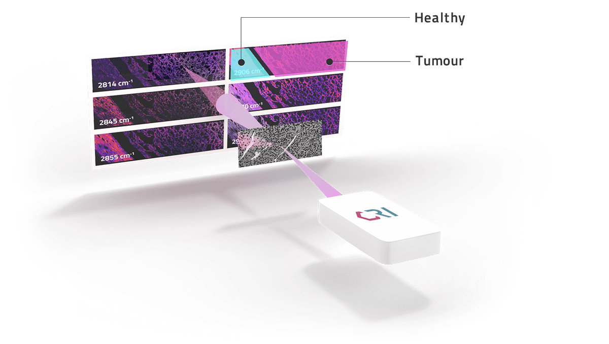
Coherent Raman Imaging has an excellent fit to the digitisation of imaging for biomedical purposes because it can produce consistent real-time, high-contrast images of a biological sample without the need for staining or labelling. By measuring the sample’s vibrational spectrum or “fingerprint”, which reflects its intrinsic molecular structure, we consistently identify tissue and cells with high-sensitivity and specificity.
ARTIFICIAL INTELLIGENCE
Chemometric data for advanced AI approaches
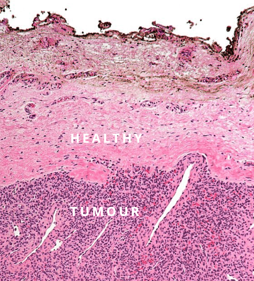
Current AI modules used in Digital Histo–pathology rely on on analyzing the shape, color, and distribution of cells in H&E stained tissue. However, this method has inherent variability due to wet chemistry staining.
Coherent Raman Imaging, beside capturing morphological information, quantifies the chemical signature of selected molecules like lipids, proteins and DNA at every point across the sample, providing a more complete picture of the tissue.
The resulting data-rich images can be leveraged with advanced AI approaches like deep convolutional neural networks, enabling consistent and accurate AI classification across multiple laboratory sites.
This capability improves the accuracy of diagnoses for diseases like tumors and provides valuable insights into cellular processes, making it an ideal solution for biotech and drug discovery applications.

Statements about future products represent management intentions, but are not to be considered commitments to any specific clinical or non-clinical capability or performance targets.


